Surface Anatomy Of Ribs - Jaypeedigital Ebook Reader : The line diverges somewhat as it descends, and lateral to it is a broad convex surface caused by the projection of the ribs beyond their angles.
bymagamalmanger-
0
Surface Anatomy Of Ribs - Jaypeedigital Ebook Reader : The line diverges somewhat as it descends, and lateral to it is a broad convex surface caused by the projection of the ribs beyond their angles.. Surface anatomy of the lungs. The costal surface is smooth and convex. Anatomy of the human body. With the upper ribs, closer to the nodule (and in the case of lower ribs, a little further from the nodule) they are curved and have a rough surface that connects them with muscles, angulus costae. Costae) are long, flat, curved bones that form the rib cage.
The line diverges somewhat as it descends, and lateral to it is a broad convex surface caused by the projection of the ribs beyond their angles. The lung is located deep to the area going from axilla to the 7th or 8th rib. Costae) are long, flat, curved bones that form the rib cage. A rib has a flat body, as you can see from the picture of the anatomy of the human rib cage. It is an atypical rib and is an important anatomical landmark.
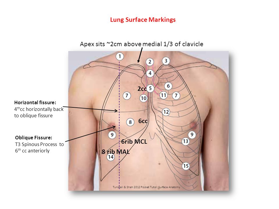
Rib Cage Wikipedia from upload.wikimedia.org However, only seven have a direct articulation with the sternum. It is one of the borders of the superior thoracic aperture. Lungs extend from about 2 cm above the clavicle down to the 6th rib in the midclavicular line and 8th rib in the midaxillary line; The costal surface is smooth and convex. Where the neck meets the body there is a roughed tubercle, with a facet for articulation with the transverse process. Surface anatomy of the back prepared by: Because rib 1 articulates with first thoracic vertebra only, there is a single facet on its head (typical ribs have two facets, as mentioned above). The border of the heart that lies inside can be traced on the front of chest wall by marking the following points:
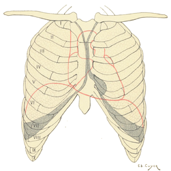
In this image, you will find right lung surface landmark diagram, superior lobe, tiv spine, 5th rib, 6th rib, middle lobe in it.
Three imaginary vertical lines on the anterior wall give you points of reference: Anatomy the rib cage is a bony structure found in the chest (thoracic cavity). The lung is located deep to the area going from axilla to the level of the 7th or 8th rib. The anterior border of the lung is formed by the convergence of the mediastinal and costal surfaces. The upper lobe is demarcated in the level of the 5th rib in the midaxillary line and 6th rib in the midclavicular line. You may also find inferior lobe, 8th rib, horizontal fissure, oblique, parietal fissure, costodiaphragmatic recess as well. The rib cage is collectively made up of long, curved. The ribs form the main structure of the thoracic cage that protects the thoracic organs. The typical rib consists of a head, neck and body: The costal surface is smooth and convex. Hilum and root of lungs 4. Ribs 11 and 12 do not have necks or tubercles and the anterior tips of their bodies lack an articular surface. The horizontal fissure (only on the right) starts at the 4th rib at the sternum and then meets the oblique fissure at the 5th rib in the midaxillary line.
The upper edge is round and the lower sharp. The heads of ribs 1, 10, 11, and 12 have a single facet for articulation with the bodies of the thoracic vertebrae. The lines cover the front, side, and back of the thorax. The ribs are curved, flat bones which form the majority of the thoracic cage. There are twelve pairs of ribs, all of which articulate with the vertebral column, while only the first seven ribs directly articulate with the sternum.
Lower Respiratory Tract Anatomy Thoracic Wall Arterial Supply Posterior Intercostal Arteries Descending Aorta Anterior Intercostal Arteries Internal Ppt Download from images.slideplayer.com The oblique fissure goes from the 6th rib midclavicular line to t3 in the back; Each vertebra therefore has a pair of superior articular facets that face posteriorly and a pair of inferior articulating facets that face anteriorly (except for t12). One facet articulates with the numerically corresponding vertebrae, and the other articulates with the vertebrae above. A thorough knowledge of thoracic anatomy is of fundamental importance to the thoracic surgeon. Surface anatomy (also called superficial anatomy and visual anatomy) is the study of the external features of the body of an animal. The rib cage is collectively made up of long, curved. The horizontal fissure (only on the right) starts at the 4th rib at the sternum and then meets the oblique fissure at the 5th rib in the midaxillary line. Contributing to their role in protecting the internal thoracic organs.
The lung is located deep to the area going from axilla to the level of the 7th or 8th rib.
It is one of the borders of the superior thoracic aperture. The costal surface is smooth and convex. Three imaginary vertical lines on the anterior wall give you points of reference: Hilum and root of lungs 4. Anatomy, types, ossification & clinical significance. However, only seven have a direct articulation with the sternum. There are twelve (12) pairs of ribs and all articulate posteriorly with the thoracic vertebrae. Anatomy of the human body. The surface projection is on the left side of the sternum opposite the left 3rd intercostal junction whereas the valve is ausculated on the left side of the sternum at the 2nd intercostal junction. The rib cage is collectively made up of long, curved. The heads of ribs 1, 10, 11, and 12 have a single facet for articulation with the bodies of the thoracic vertebrae. The anterior surface of the typical ribs is flat and smooth, its posterior rough for the attachment of the ligament of the neck, and perforated by numerous foramina. A rib has a flat body, as you can see from the picture of the anatomy of the human rib cage.
With the upper ribs, closer to the nodule (and in the case of lower ribs, a little further from the nodule) they are curved and have a rough surface that connects them with muscles, angulus costae. The first rib is the most superior of the twelve ribs. The junction between the body of the sternum and the xiphoid process is on the level of the tenth thoracic vertebra. A thorough knowledge of thoracic anatomy is of fundamental importance to the thoracic surgeon. There are twelve (12) pairs of ribs and all articulate posteriorly with the thoracic vertebrae.
Surface Anatomy Of The Thorax Youtube from i.ytimg.com With the upper ribs, closer to the nodule (and in the case of lower ribs, a little further from the nodule) they are curved and have a rough surface that connects them with muscles, angulus costae. The ribs are curved, flat bones which form the majority of the thoracic cage. The lung is located deep to the area going from axilla to the level of the 7th or 8th rib. Because rib 1 articulates with first thoracic vertebra only, there is a single facet on its head (typical ribs have two facets, as mentioned above). To reduce the surface tension of alveoli mainly during expiration, thus reduces the work of lung inflation. Mark a point 1cm from the right sterna line along the upper border of 3 rd right costal cartilage. Ribs the ribs partially enclose and protect the chest cavity, where many vital organs (including the heart and the lungs) are located. The lung is located deep to the area going from axilla to the 7th or 8th rib.
In birds this is termed topography.
The junction between the body of the sternum and the xiphoid process is on the level of the tenth thoracic vertebra. The lung is located deep to the area going from axilla to the level of the 7th or 8th rib. The upper lobe is demarcated in the level of the 5th rib in the midaxillary line and 6th rib in the midclavicular line. The typical rib consists of a head, neck and body: Surface projections of the major organs of the trunk, using the vertebral column and rib cage as main reference points of surface anatomy. With the upper ribs, closer to the nodule (and in the case of lower ribs, a little further from the nodule) they are curved and have a rough surface that connects them with muscles, angulus costae. A thorough knowledge of thoracic anatomy is of fundamental importance to the thoracic surgeon. In birds this is termed topography. The lines cover the front, side, and back of the thorax. Each vertebra therefore has a pair of superior articular facets that face posteriorly and a pair of inferior articulating facets that face anteriorly (except for t12). Anatomy of the human body. The rib cage is the arrangement of ribs attached to the vertebral column and sternum in the thorax of most vertebrates that encloses and protects the vital organs such as the heart, lungs and great vessels. The oblique fissure goes from the 6th rib midclavicular line to t3 in the back;
The line diverges somewhat as it descends, and lateral to it is a broad convex surface caused by the projection of the ribs beyond their angles anatomy of ribs. Hilum and root of lungs 4.